Are you looking for an essay on ‘Ultra Structure of the Cell Wall’? Find paragraphs, long and short essays on ‘Ultra Structure of the Cell Wall’ especially written for school and college students.
Essay on Cell Wall
Essay Contents:
- Essay on the Introduction to Cell Wall
- Essay on the Plant Cell Wall
- Essay on the Algal Cell Wall
- Essay on the Fungal Cell Wall
- Essay on the Protists Cell Wall
- Essay on the Bacterial Cell Wall Structure
- Essay on the Cell Wall Organization of Acid Fast Bacteria
- Essay on the Archaea Bacterial Cell Wall
1. Essay on the Introduction to Cell Wall:
A cell wall is the outer most rigid layer of the cell, located external o the cell membrane which provide the cell, structural support, protection, and act a a filtering mechanism. In multi-cellular organisms, it permits the organism to build and to hold its shape (morphogenesis). The cell wall also limits the entry of large molecules that may be toxic to the cell. It further permits the creation of a stable osmotic environment by preventing osmotic lysis and help to retain water. It is found in plants, bacteria, archaea fungi, and algae, while it is absent in animal and most protists.
The structural component of the cell wall varies according to the type of cells. In plants, the cell wall is made up of carbohydrate polymer, cellulose and in bacteria it is of peptidoglycan, while in fungi, the cell wall is made up of chitin.
2. Essay on the Plant Cell Wall:
The cell wall of plant is one of the most important distinguishing features. In addition to the protection of the intracellular contents, it gives rigidity to the plant, provides a porous medium for the circulation and distribution of water, minerals, and other nutrients, and houses a number of specialized molecules that regulate growth and protect the plant from diseases. The thickness, as well as the composition and organization of the cell wall can vary significantly.
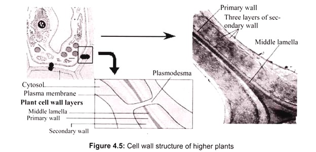
The cell wall has three major regions:
i. Middle Lamella:
It is pectin layer which cements the cell wall of two adjoining cells together. Plants need this lamella to give them stability so that they can form plasmodesmata between the cells. It is the first layer which is deposited at the time of cytokinesis. The Middle lamella is mainly made up of calcium and magnesium pectate.
ii. Primary Wall:
It is deposited by cells before and during active growth. The actual content of the wall components varies from species to species with the age. All plant cells have a middle lamella and primary wall.
iii. Secondary Wall:
Some cells deposit additional layers inside the primary wall. This occurs especially after the growth stops or when the cell begins to differentiate. This wall is mainly for support and is comprised primarily of cellulose and lignin. It has distinct layers S1, S2 and S3, which differ in the orientation, or direction of the cellulose microfibrils.
Chemical Composition of Plant Cell Wall:
Cellulose:
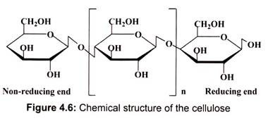
Cellulose is the major component of plant cell walls. It is an un-branched polymer with about ten thousand glucose units per chain. Hydroxyl groups (-OH) project out from each chain, forming hydrogen bonds with neighboring chains which creates a rigid cross-linking between the chains, making cellulose as the strong support material. Despite the combined strength of cellulose, it is fully permeable to water and solutes which makes it ideal for allowing water and solutes into and out of the cell. It is the most abundant organic substance found in the living world and has been estimated to be more than half of the total organic carbon available on the planet (Fig. 4.5).
Cellulose readily forms hydrogen bonds with itself (intra-molecular H-bonds) and with other cellulose chains (inter-molecular H-bonds). A cellulose chain forms hydrogen bonds with about 36 other chains in order to yield a micro-fibril. This is somewhat analogous to the formation of a thick rope from thin fibers. Micro-fibrils are 5-12 nm wide and offer the wall strength (Fig. 4.6). They have a tensile strength equivalent to steel. Some regions of the micro-fibrils are highly crystalline while others are more “amorphous”.
Hemicelluloses:
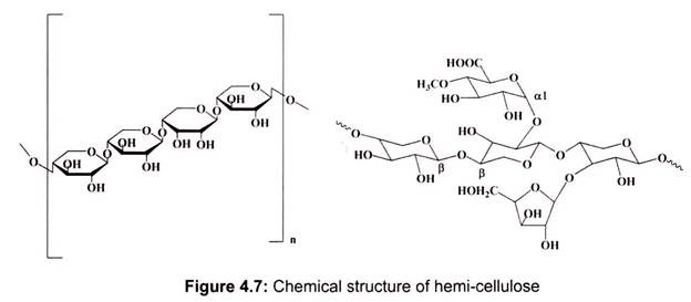
They are branched polysaccharides structurally homolgous to cellulose because they have a backbone composed of 1, 4-linked β-D-hexosyl residues. The predominant hemi-cellulose in many primary walls is xyloglucan. Other hemicelluloses found in primary and secondary walls include glucuronoxylan, arabinoxylan, glucomannan, and galactomannan. They are characterized by being soluble in strong alkali.
The main feature of this group is that they do not aggregate with themselves – in other words, they do not form micro-fibrils. However, they form hydrogen bonds with cellulose and hence the reason they are called “cross-linking glycans”. Hemi-cellulose molecules are often branched. Like the pectic compounds, hemi- cellulose molecules are highly hydrophilic. They become exclusively hydrated and form gels. Hemi-cellulose is abundant in primary walls and also found in secondary walls (Fig. 4.7).
Pectin:
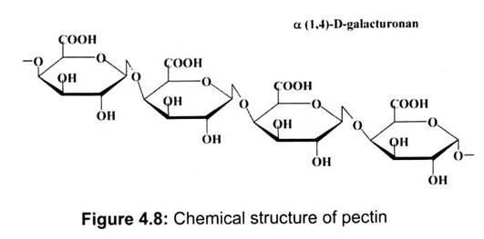
It is a family of complex polysaccharides that contain 1, 4-linked α-D-galacturonic acid. There are three represent classes of pectic polysaccharides. They are comparatively less hydrated than pectic acid but soluble in hot water. It is another major component of middle lamella but also found in primary walls (Fig. 4.8). This is made up of a modified sugar, galacturonic acid and in the plant the carboxyl groups are esterified with methyl (CH3) groups.
Divalent cations, like calcium, also form cross-linkages amongst adjacent polymers creating a gel. Pectic polysaccharides can also be cross-linked by dihydrocinnamic or diferulic acids. The pectic polysaccharides serve a variety of functions including determining wall porosity, a charged wall surface for cell-cell adhesion (middle lamella), cell-cell recognition, pathogen recognition and others.
Proteins:
Wall proteins are typically glycol-proteins (polypeptide backbone with carbohydrate side chains). The proteins are particularly rich in the amino acids hydroxyl-proline (hydroxyproline-rich glycoprotein, HPRG), and glycine (glycine-rich protein, GRP). These proteins form rods (HRGP, PRP) or beta-pleated sheets (GRP). HRGP (Extensin) is induced by wounding and pathogen attack. Protein appears to be cross-linked to pectic substances and may have sites for lignifications. The proteins may serve as the scaffolding used to construct the other wall components.
Lignin:
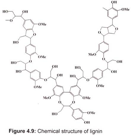
Lignin is a complex polymer of phenyl-propane units, which are cross-linked to each other with a variety of different chemical bonds. This complexity has thus far proven as resistant to microbial degradation. Nonetheless, some organisms, particularly fungi, have been identified with the necessary enzymes that break up lignin apart.
Lignin is a major chemical compound present in the cell walls of tracheids, xylary fibres and sclereids of plants. Lignin polymers are mostly made up of phenyl-propanoid units linked to each other through different kinds of bonds. Lignin polymers are probably network structures with molecular weights on the order of 10,000 amu. Lignin is the most abundant organic material on earth after cellulose.
It is believed that lignin gives wood its stiffness and improves water transport (Fig. 4.9). Lignin is thought to act as a kind of glue in the plant cell walls and give plants very effective protection against parasite attack. Lignin makes up about one-quarter to one-third of the mass of dry wood. In the chemical pulping process, lignin is removed from wood pulp before it is turned into paper, and the extracted lignin is used as a binder in particle board, adhesive for linoleum, and raw material for processing into chemicals (such as DMSO and vanillin). The type of lignin (such as lingo-sulfonates and kraft lignins) used in industry depends upon the method that was used to extract it.
Suberin and Cutin:
Suberin is fairly hydrophobic and its main function is to prevent water from penetrating the tissue. A variety of lipids are associated with the wall for strength and water-proofing. Cutin is a waxy polymer that is the main components of the plant cuticle which covers all aerial surfaces of plants.
Water:
The cell wall is largely hydrated and comprised of between 75—80% water. This is responsible for some of the wall properties. For example, hydrated walls exhibit greater flexibility and extensibility than non-hydrated walls.
3. Essay on the Algal Cell Wall:
Algae are a large and diverse class of simple, typically autotrophic organisms. The cell wall composition and structure of algae differs significantly from that of higher plants. Neither in composition nor in biosynthesis do they have any common features with the cell walls of plants.
In many classes of algae cellulose is the main structural element of the wall, though remarkable variations of the fibrillary structure may exist. In some classes of algae there are only dispersed textures, while in others (especially many Chlorophyta-species) a higher degree of organization (layers of parallel micro-fibrils) is present. No clear difference between primary and secondary cell wall exists in most algae.
Manosyl:
Manosyl constitutes the main structural elements in a number of marine green algae (Codium, Dasycladus, Acetabularia, etc.) as well as in the walls of some red algae (Porphyra, Bangia). They are linear and the mannosyl residues are (1-4) glycosidically linked.
Xylanes:
They are the polymers where the β-D-xylosyl residues are linked through (1-3) and (1-4) glycosidic bonds. In contrast to other polymers, xylans are partially ramified. In species with xylan-containing walls there exists a layered structure and an orientation of the microfilaments. They contain mostly linear polymers.
Alginic Acid:
The cell walls of Phaeophyta (brown algae) mainly consist of alginic acid and its salts, the alginates, consist exclusively of uronic acids: mannuronic acid and beta-L- glucuronic acid in varying ratios along with small amounts of β-D-glucuronic acid. It is a linear copolymer consisting mainly of residues of β (1, 4)-linked D-mannuronic acid and α -1,-4-linked L-glucuronic acid. These monomers are often arranged in homo-polymeric blocks separated by regions approximating an alternating sequence of the two acid monomers (Fig.4.10).
The alginates of brown algae exist both within the cell wall and in the intercellular substance. Their part in the cell wall may be as high as 40 per cent of the dry matter. They have a high affinity for divalent cations (calcium, strontium, barium, magnesium) and a tendency to gel.
Sulfonated Polysaccharides:
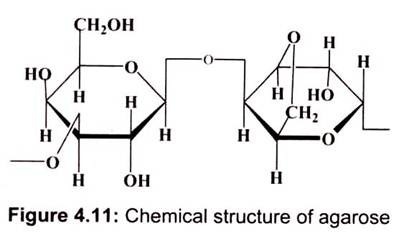
They occur partially in the cell wall itself and partially in the intercellular substance. Sulfonated galactanes are typical for many red algae, depending on their origin they are called agarose, carrageenan, porphyran, furcelleran and funoran. Chemically they are polysaccharides whose monomers are esterified to sulfuric acid residues and moreover they are partially methylated.
Carrageenan and furcelleran contain exclusively D-compounds. Agar, whose basic unit is agarose, is obtained mainly from Gelidium and Gracillaria, both genera of red algae. The extraordinary binding types of agarose and carrageenan lead to specific tertiary structures.
Silicon:
It is one of the main components of the diatom shell. It also occurs in the cell walls of other groups of algae. Silicon is a cell wall component in some brown algae and in the green algae Hydrodictyon. Diatoms take up silicon as silicate. Chemically, agarose is a polysaccharide, whose monomeric unit is a disaccharide of D- galactose and 3, 6-anhydro-L-galactopyranose (fig. 4.11).
Calcium:
Calcium seems to be common in species of tropical and marine waters. Some species participate in reef formation.
Calcium is always deposited as calcium carbonate and occurs in two different crystalline states:
a. Calcite and
b. Aragonite.
Calcite is produced in the walls of some groups of red algae and in Charophyceae, while aragonite is produced by some green (Acetabularia, etc.), brown and red algae. Both states do not occur simultaneously in one species.
4. Essay on the Fungal Cell Wall:
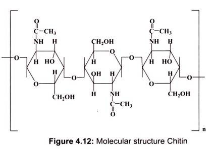
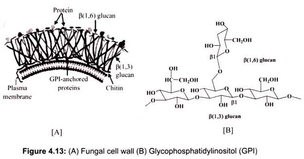
The fungal cell wall is a complex structure composed typically of chitin (fig. 4.12), 1,3- β- and 1,6-β-glucan, mannan and proteins, although the wall composition frequently varies markedly between species of fungi, there is evidence of extensive cross-linking between these components. Fungi have chitin cell walls, which make them more like animals than plants. Conversely, the water molds have cellulose cell walls that make them more plantlike, so they have been separated from the fungi in modern classification.
Approximately 80% of the wall consists of polysaccharides. Most fungi have a fibrillar structure buildup of chitin chitosan (Zygomycota only), and β-glucans, and a variety of hetero-polysaccharides (Fig. 4.13). The fibres are contained in a complex gel-like matrix. Proteins constitute a small fraction of wall material, rarely more than 20%, and often as glycoprotein. Not all proteins have a structural role to play. Hydrophobins are expressed constitutively and become bound in the wall, when the hyphae emerge in air. Lipids are found in walls, usually in very small concentrations.
Along with hydrophobins, lipids and waxes appear to function in the process of control over movement of water, especially the prevention of desiccation of cells. Walls also contain a range of other minor components, including pigments and salts. The pigments, melanin is particularly intriguing. Melanin, important for protecting the hyphae and spores from UV stress, is essential for pathogenesis and attachment to surfaces of emerging hyphae from spores.
The constituents of cell walls are synthesized in the cytoplasm, linked in the walls at the hyphal tip, polymerized and cross-linked in the wall matrix. Chitin and the glucans are synthesized at the plasma membrane by the enzymes embedded in the membrane itself. Nucleotide sugar precursors are accepted from the cytoplasm, linked and passed to the wall. The wall glycol-proteins are synthesized in the endoplasmic reticulum carried through the Golgi to the plasma membrane, where vesicles release the glycoprotein to the wall. Enzymes cross- linking fibrils in the wall are released through the plasma membrane.
Hydrophobins:
Hydrophobins constitute up to 10% of total wall protein. The amphipathic structure (having hydrophobic and hydrophilic domains) provides the molecules with an extraordinary potential array of functions. This construction reduces movement of water through the wall of the hyphae providing some protection from desiccation. It may also increase the strength of the wall. The exposed hydrophobic domain enables attachment to hydrophobic surfaces. The hyphae can also attach to hydrophobic (e.g. waxy) plant surfaces, enabling attachment of spores prior to formation of appressoria. The attachment properties make hydrophobins extremely important in morphogenic development.
Glomalin:
Some members of the Glomeromycota produce a putative glycoprotein in the walls. This compound has been called glomalin. Glomalin is important because of its possible correlation with aggregation of soil particles and the purported resilience of the molecule.
5. Essay on the Protists Cell Wall:
Protists are all eukaryote$ (cell with nuclei). All protists live in moist surrounding, unicellular, while some are multi-cellular. Some are heterotrophs (cannot make their own food; have to eat something) some are autotrophs (make their own food) some cannot move (sessile), while others can move. These are divided into three categories, i.e., animal-like, plant-like and fungus-like protists.
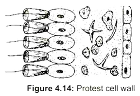
The cells of many protists are surrounded by a filamentous layer of oligosaccharides. This forms the cell coat. It is in fact a part of the cell membrane. In many cases the cell coat becomes deposited with salts of calcium and silica (Fig.4.14). The cell coat mainly provides protection to the cell from infection by pathogens. It also helps in the recognition of similar cells.
6. Essay on the Bacterial Cell Wall Structure:
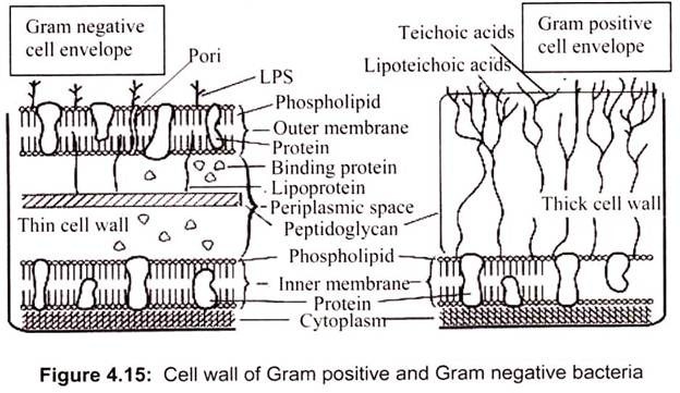
The cell wall lies outside the cell membrane. The rigid peptidoglycan is important in defining the shape of the cell and giving the cell a mechanical strength. Bacteria may be conveniently divided into two groups, depending upon their ability to retain a crystal violet-iodine dye complex when cells are treated with acetone or alcohol (Gram reaction, named after Christian Gram). This reaction, however, reveals fundamental differences in the structure of bacteria. Cells with many layers of peptidoglycan can retain a crystal violet-iodine complex when treated with acetone. These are called Gram-positive bacteria and appear blue-black or purple when stained using Gram’s method (Fig. 4.15). Gram-negative bacteria have only one or two layers of peptidoglycan and cannot retain the crystal violet-iodine complex. They require counterstaining with another dye (safranin) to be seen using Gram’s method.
Gram negative bacteria have a much thinner cell wall. The wall is high in lipid content and low in peptidoglycan. The crystal-violate escapes from cell when the decolorizer is added because of the lack of peptidoglycan. The outer membrane of gram negative bacteria is composed of a high concentration of lipids, polysaccharides and proteins. Most Gram- positive bacteria have a relatively thick (about 20 to 80 nm), continuous cell wall, which is composed largely of peptidoglycan (also known as mucopeptide or murein). In thick cell walls, other cell wall polymers such as the teichoic acids, polysaccharides, and peptidoglycolipids are covalently attached to the peptidoglycan.
In contrast, the peptidoglycan layer in Gram-negative bacteria is thin (about 5 to 10 nm thick); in E coli, the peptidoglycan is probably only a monolayer thick. Outside the peptidoglycan layer in the Gram-negative envelope, there is an outer membrane structure (about 7.5 to 10 nm thick). In most Gram- negative bacteria, this membrane structure is anchored non-covalently to lipoprotein molecules, which in turn, are covalently linked to the peptidoglycan.
The lipo-polysaccharides of the Gram-negative cell envelope form outer part of the outer membrane structure:
(a) Peptidoglycans:
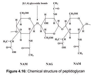
Both Gram-positive and Gram-negative bacteria possess cell wall peptidoglycans, which confer the characteristic cell shape and provide the cell with mechanical protection. Peptidoglycans are unique to prokaryotic organisms and consist of a glycan backbone of muramic acid and glucosamine (both N-acetylated; N- acetyl muramic acid (NAM) and N- acetyl glucosamine (NAG)), and peptide chains highly cross-linked with bridges in Gram-positive bacteria (Staphylococcus aureus) or partially cross-linked in Gram- negative bacteria (E. coli).
The main function of peptidoglycan is to preserve cell integrity by withstanding the turgor pressure inside the cell. Inhibition of peptidoglycan biosynthesis by mutations or antibiotics such as penicillin or degradation by lysozyme in growing cells results in cell lysis (Fig. 4.16). Peptidoglycan contributes to the maintenance of a defined cell shape (rod or sphere) and serves as a scaffold for anchoring other cell envelope components such as proteins and teichoic acids. However, peptidoglycan is found absent in some bacteria (.Mycoplasma sp., Planctomyces, Rickettsia and Chlamidiae).
(b) Teichoic Acids:
Membrane associated teichoic acids (lipoteichoic acids) are polymers of amphiphilic glyco- phosphates with the lipophilic glycolipid and anchored in the cytoplasmic membrane. They are antigenic, cytotoxic and adhesions (Streptococcus pyogenes). Teichoic acids are polyol phosphate polymers, with either ribitol or glycerol linked by phosphordiester bonds bearing a strong negative charge. They are covalently linked to the peptidoglycan in some Gram- positive bacteria. They are strongly antigenic, but are generally absent in Gram-negative bacteria. Teichoic acids are covalently linked to the peptidoglycan.( Fig. 4.17). These highly negatively charged polymers of the bacterial wall can serve as a cation-sequestering mechanism.
(c) Lipo-Polysaccharides (LPS):
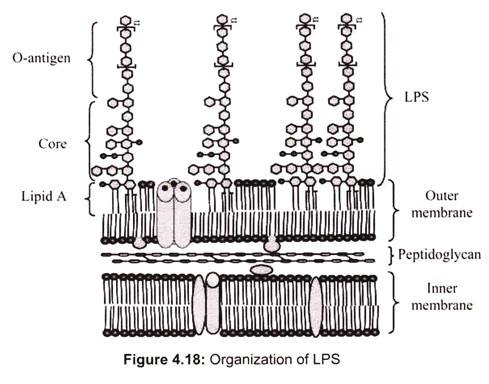
One of the major components of the outer membrane of Gram-negative bacteria is lipo- polysaccharide (endo-toxin), a complex molecule consisting of a lipid anchor, a polysaccharide core, and chains of carbohydrates. Sugars in the polysaccharide chains confer serologic specificity. So far, only one Gram-positive organism, Listeria monocytogenes, has been found to contain an authentic LPS.
Endo-toxins possess an array of powerful biologic activities and play an important role in the pathogenesis of many Gram-negative bacterial infections. In addition to causing endo-toxic shock, LPS is pyrogenic, can activate macrophages and complement, mitogenic for B lymphocytes, induces interferon production, causes tissue necrosis and tumor regression and has adjuvant properties.
Usually, the LPS molecules have three regions: the lipid structure required for insertion in the outer leaflet of the outer membrane bilayer; a covalently attached core composed of 2- keto-3deoxyoctonic acid (KDO), heptose, ethanolamine, N-acetyl-glucosamine, glucose, and galactose; and polysaccharide chains linked to the core. The polysaccharide chains constitute the O-antigens of the Gram-negative bacteria, and the individual monosaccharide constituents confer serologic specificity on these components. LPS and phospholipids help confer asymmetry to the outer membrane of the Gram-negative bacteria, with the hydrophilic polysaccharide chains outermost. Each LPS is held in the outer membrane by relatively weak cohesive forces (ionic and hydrophobic interactions) and can be dissociated from the cell surface by action of surface-active agents (Fig. 4.18).
7. Essay on the Cell Wall Organization of Acid Fast Bacteria:
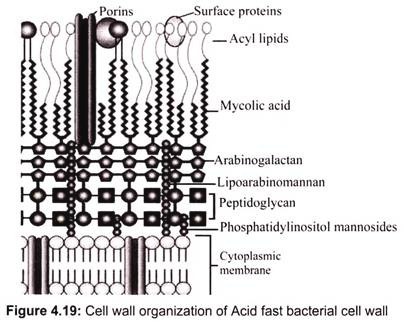
Acid-fast bacteria are gram-positive. Acid fast bacterial cell wall is composed of a thin, inner layer of peptidoglycan and large amount of glycolipids, such as mycolic acid, arabinogalac- tan-lipid complex, and lipoarabinomannan. It is approximately 60 percent lipid and contains much less peptidoglycan (Fig. 4.19).
The lipids make acid-fast organisms impermeable to most other stains and protect them from acids and alkalis. During the acid-fast staining procedure, the acid-fast cell wall enables the bacterium to resist de-colorization with acid alcohol and retain the original stain carbol fuchsin. Like the outer membrane of the gram-negative cell wall, porins are required to transport small hydrophilic molecules through the outer membrane of the acid-fast cell wall.
There are far fewer porins in the acid-fast cell wall compared to the gram-negative cell wall and the pores are much longer. The peptidoglycan prevents osmotic lysis and the mycolic acids and other glycol-lipids impede the entry of chemicals which cause the organisms to grow slowly and be more resistant to chemical agents and lysosomal components of phagocytes than most bacteria. This is thought to contribute significantly to the lower permeability of acid-fast bacteria.
8. Essay on the Archaea Bacterial Cell Wall:
Archaeal cell walls lack peptidoglycan, except one group of methanogens. In this, the peptidoglycan is a modified form, different from that found in bacteria.
There are four types of cell wall currently known among the Archaea:
i. Pseudo-peptidoglycan (also called pseudomurein) is the main structural component of cell wall of some methanogens (Methanobacterium and Methanothermus). Pseudo- peptidoglycan consists of polymer chains of glycan cross-linked with short peptides. However, unlike peptidoglycan, the sugar N-acetylmuramic acid is replaced by N- acetyltalosaminuronic acid, and the two sugars are bonded with a β,1-3 glycosidic linkage instead of β,1-4, also cross-linking peptides are L-amino acids rather than D-amino acids as in bacteria.
ii. The second type of cell wall is composed entirely of a thick layer of polysaccharides, which may be sulfated in case of Methanosarcina and Halococcus.
iii. Third type of Archaeal cell wall consists of glycoprotein, which occurs in the hyperthermophiles, Halobacterium, and some methanogens. In Halobacterium, the proteins in the wall have a high content of acidic amino acids, giving the wall an overall negative charge and thrive only under conditions with high salinity since structure is stabilized by the presence of large quantities of positive sodium ions that neutralize the charge.
iv. In the fourth type, the wall is composed of only of surface-layer proteins, known as an S- layer. S-layers are monomolecular crystalline arrays of proteinaceous subunits and are common in bacteria, where they serve as either the sole cell-wall component or an outer layer in conjunction with peptidoglycan and murein. Most Archaea are Gram-negative, though at least one Gram-positive member is known.
Methanomicrobium and Desulfurococcus are belonging to this category.
